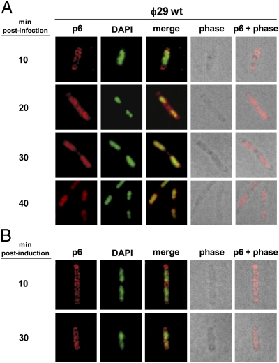
Fig. 1.
Subcellular localization of protein p6 in ϕ29-infected and noninfected cells. B. subtilis 110NA and IH-04 (expressing p6) cells were grown at 37 °C in LB medium supplemented with 5 mM MgSO4. At an OD600 of 0.45–0.5, the 110NA culture was infected with wild-type phage ϕ29 at a multiplicity of infection (MOI) of 5 (A), and IPTG was added to a final concentration of 1 mM to the IH-04 culture (B). Samples were withdrawn at the indicated times post infection or after IPTG addition and subjected to IF microscopy using polyclonal antibodies against p6. For clarity, p6 and DAPI fluorescent signals are false-colored red and green, respectively.

