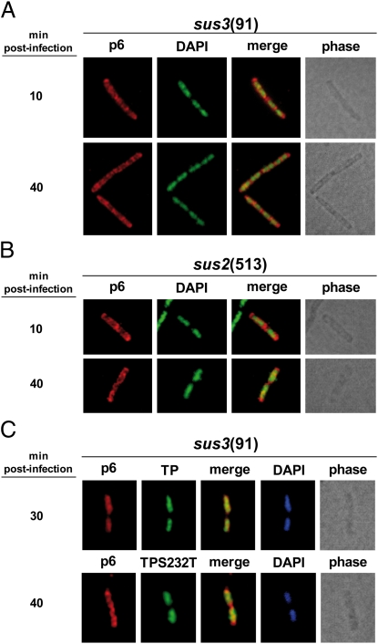
Fig. 3.
The nucleoidal localization of protein p6 depends on viral DNA replication. (A and B) B. subtilis 110NA cells were grown at 37 °C in LB medium supplemented with 5 mM MgSO4. At an OD600 of 0.45–0.5, the cultures were infected with sus3(91) or sus2(513) mutant phages at a MOI of 5. Samples were harvested at the indicated times post infection and processed for IF microscopy using polyclonal antibodies against p6. For clarity, p6 and DAPI fluorescent signals are false-colored red and green, respectively. (C) Strains IH-16 (expressing TP wild type) and IH-18 (expressing TP-S232T mutant) were grown at 37 °C in LB medium supplemented with 5 mM MgSO4. At an OD600 of 0.45–0.5, the cultures were infected with sus3(91) mutant phage at a MOI of 5, and 10 μM IPTG was added to the medium 10 min later. Samples were withdrawn 30 or 40 min post infection and subjected to IF microscopy using polyclonal antibodies against both proteins, p6 and TP.

