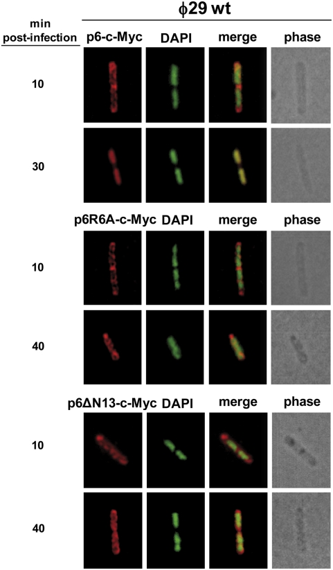
Fig. 4.
Protein p6 mutants in DNA binding are not distributed at the bacterial nucleoid. B. subtilis strains IH-10 (expressing p6-c-Myc), IH-12 (expressing p6R6A-c-Myc), and IH-14 (expressing p6ΔN13-c-Myc) were grown at 37 °C in LB medium containing 5 mM MgSO4. At an OD600 of 0.45–0.5, cells were infected with wild-type phage ϕ29 at a MOI of 5, and 50 μM IPTG was added to the medium 5 min later. Samples were withdrawn at the indicated times post infection and subjected to IF microscopy using polyclonal antibodies against p6. For clarity, p6 and DAPI fluorescent signals are false-colored red and green, respectively.

