Figure 2.
the structure of ArnA
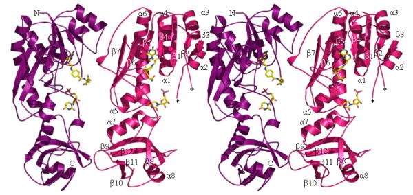
2a The structure of the formyltransferase domain. The protein is represented as ribbons and the secondary structure elements are numbered as the text. A loop (residues N35-A40) that is missing in the structure is labeled *. The two subdomains are visible, the N-terminal subdomain is at the top of the figure and extends to the uridine ring, the C-terminal subdomain sits below the ring. The dimer found in the crystal is shown. The decarboxylase domain is attached to the C-terminus (labeled C).
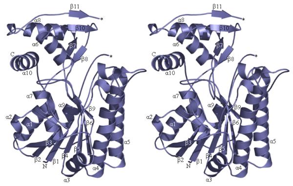
2b The structure of the decarboxylase domain. The protein is represented as ribbons and the secondary structure elements are numbered. A loop (residues V604-D615) that is missing in the structure is labeled *. The monomeric unit is shown. The N-terminus of this domain (labeled N) is attached to the formyltransferase domain.
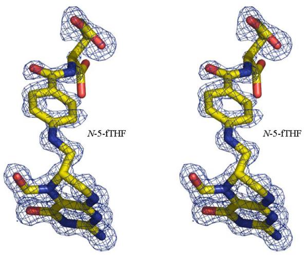
2c The unbiased Fo-Fc electron density map for N-5-fTHF found in the formyltransferase domain. The molecules are shown in stick with atoms colored; carbon yellow, nitrogen blue, oxygen red and phosphorous purple. The map is contoured at 3σ (0.22eÅ−3)
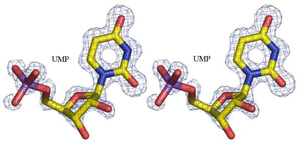
2d The unbiased Fo-Fc electron density map for UMP found in the formyltransferase domain. Contouring and atomic color are the same as Figure 2c.


