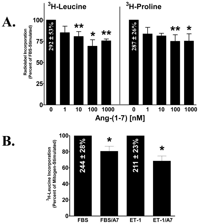
Figure 3. Inhibition of protein and collagen synthesis by Ang-(1-7) in cardiac fibroblasts.
Cardiac fibroblasts were deprived of serum for 24 h and subsequently treated for 48 h with various concentrations of Ang-(1-7), in the presence of 1% FBS or 10 nM ET-1. Protein and collagen was detected by 3H-leucine and 3H-proline incorporation, respectively, which were added during the last 24 h of the treatment period. Data are presented as the percentage of mitogen-stimulated growth, in the absence of Ang-(1-7). n = 4 – 6, in triplicate. *denotes p < 0.05 and **denotes p < 0.01 compared to mitogen alone.

