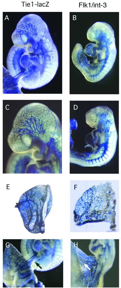
Figure 2.
Embryo whole-mounts composite. Control Tie1-lacZ (A and C) and Flk1/int-3 (B and D) embryos at 9.5 dpc were processed for whole-mount β-galactosidase staining to visualize the endothelial cells. At 9.5 dpc, vasculature in mutant embryos appears to be more restricted and disorganized. Yolk-sac vasculature is shown for Tie1-lacZ (E) and Flk1/int-3 (F). Analysis of vessels in the umbilical cord (arrow) is shown for Tie1-lacZ (G) and Flk1/int-3 (H).

