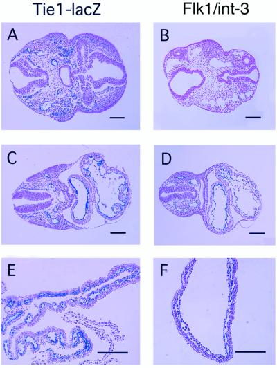
Figure 3.
Embryo sections composite. β-Galactosidase staining of histological sections of Tie1-lacZ (A, C, and E) and Flk1/int-3 embryos (B, D, and F) at 9 dpc. A and B show cross sections through the brain. C and D show cross sections through the spinal cord, cardinal vessels, and heart. E and F show cross sections through yolk sacs. In Flk1/int-3 embryos, enlarged and collapsed vessels are observed, large areas of necrosis are seen, and fewer β-galactosidase-staining cells are visible (B and D). (Bars = 1 mm.)

