
Figure 4. FTS decreases secretion of the immunosuppressive cytokine TGF-β from GL261 glioma cells and increases the proliferative and cytotoxic capacities of CTLs in vitro as well as accumulation of CTLs in the tumors in vivo.
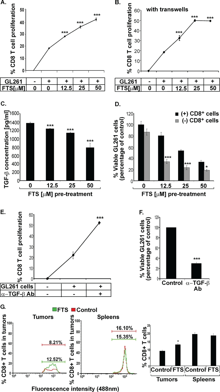
(A) GL261 tumor cells were treated in vitro for 24 hours with FTS or CD8 or both, as described in Results. The cells were then washed thoroughly, and CFSE-labeled CD8+ T cells, (isolated from FTS- or vehicle-pretreated GL261 tumor-bearing mouse splenocytes) were added and cocultured with the FTS-pretreated GL261 cells for 96 hours. The rate of CTL proliferation was measured by flow cytometry. Statistical analysis of the results is presented as means ± SEM (n=8). ***, p<0.001 compared with vehicle-treated mice. (B) The experiment was performed as in A, except that the CD8+ T cells were now separated from the GL261 tumor cells by a transwell, preventing cell passage. The rate of CD8+ T cell proliferation was measured by flow cytometry. Statistical analysis of the results is presented as means ± SEM (n=8). ***, p<0.001 compared with vehicle-treated mice. (C) GL261 tumor cells were treated with the indicated doses of FTS or with vehicle (control) for 24 hours and then assayed for TGF-β (see Methods). The ELISA results are shown (means ± SEM, n=8). ***, p<0.001 compared with vehicle-treated control cells. (D) Isolated CD8+ T cells were cocultured with FTS-pretreated GL261 cells for 96 hours and their proliferation was analyzed, as described in Methods. Numbers of viable CTLs are presented as means ± SEM (n=8). *, p<0.05, ***, p<0.001 compared with vehicle-treated cells. (E-F) GL261 tumor cells were incubated with CFSE-labeled-CD8+ T cells, with or without TGF-β-blocking anti-TGF-β Ab, for 96 hours. The rate of CD8+ T cell proliferation was measured by flow cytometry (E), and viable GL261 cells were counted (F). Statistical analysis of the results is presented as means ± SEM (n=5). ***, p<0.001 compared with cells not treated with anti-TGF-β Ab. (G) C57bl/6 mice implanted s.c. with GL261 tumor cells were divided into two groups for treatment with FTS (n=10) or vehicle (n=10), as described in Methods. The mice were killed 21 days after the cells were implanted and their tumors and spleens were assayed for CD8+ T cells by flow cytometry (left and middle). Statistical analysis of the flow cytometry results (n=10) is presented (right). *, p<0.05 compared with vehicle-treated controls.