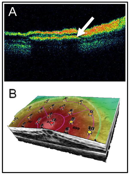
Figure 3.

Use of advanced imaging methodology to ascertain implant positioning and retinal integrity. A) using optical coherence tomography (OCT), the cross-sectional position of the implanted microelectrode array (arrow) can be viewed in subretinal space. B) Three dimensional combined OCT with retinal microperimtery allows for simultaneous assessment of structure and function at specific points of retina. The resulting topographic map (values indicate luminance levels detected in decibels; dB) could potentially be used for post-operative evaluation as well as pre-operative assessment of candidate implant locations. Image generated using an OPKO/OTI combined optical coherence tomography and scanning laser ophthalmoscope with microperimetry feature (Opko Health Inc. Miami, FLA).
