Fig. 2.
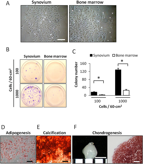
Figs. 2-A through 2-F Synovial mesenchymal stem cells have high proliferation capacity and multipotentiality. Synovium refers to synovial mesenchymal stem cells, and bone marrow refers to bone-marrow mesenchymal stem cells. Fig. 2-A Histological appearance of synovial mesenchymal stem cells (left) and bone-marrow mesenchymal stem cells (right) at passage 3. Both groups formed monolayers of spindle-shaped cells that adhered to plastic culture dishes. Scale bar indicates 200 μm. Fig. 2-B Colony formation of synovial mesenchymal stem cells and bone-marrow mesenchymal stem cells at passage 3. Nucleated cells from synovium and bone marrow were plated at 100 and 1000 cells per 60-cm2 dish and cultured for fourteen days (n = 5 cultures each). Culture dishes stained with crystal violet are shown. Fig. 2-C Graph showing the number of colonies (>2 mm) per dish at 100 or 1000 cells per 60 cm2. *P < 0.01. Fig. 2-D Adipogenesis. Adipocyte colonies were stained with oil red O. Scale bar represents 200 μm. Fig. 2-E Calcification. Calcified colonies were stained with alizarin red. Scale bar represents 500 μm. Fig. 2-F Chondrogenesis. Gross photograph of a pellet (left). Scale bar represents 1 mm. Histological section of the pellet stained with safranin O (right). Scale bar represents 100 μm.

