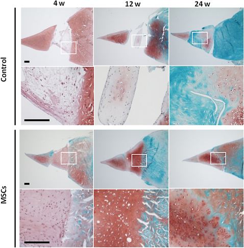
Synovial mesenchymal stem cells (MSCs) promote the meniscal regeneration (histological observation).
Fig. 5-A.
Low and high-power images of representative sections of regenerated meniscus stained with safranin O at four, twelve, and twenty-four weeks after implantation of synovial mesenchymal stem cells. The inset shows the area seen at higher magnification in the photomicrograph below. Scale bars represent 200 μm.
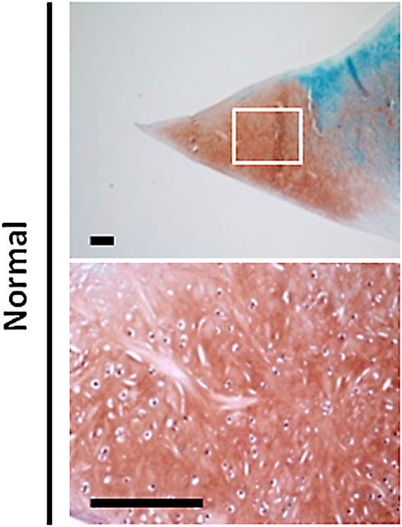
Fig. 5-B.
Low and high-power representative images of the normal meniscus stained with safranin O. Scale bars represent 200 μm.
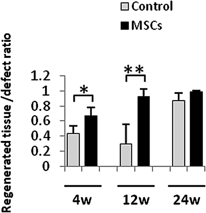
Fig. 5-C.
Regenerated tissue-to-defect ratios are displayed as the mean and the standard deviation for synovial mesenchymal stem cell (MSCs) and control groups at each end point. *The difference between the groups was significant (p < 0.05). **The difference between the groups was significant (p < 0.01).
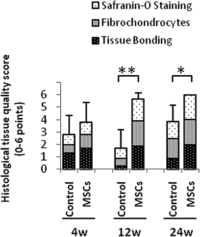
Fig. 5-D.
Results of the histological scoring system for regenerated meniscus. The scores are displayed as the mean and the standard deviation. *The difference between the groups was significant (p < 0.05). **The difference between the groups was significant (p < 0.01).


