Figure 1.

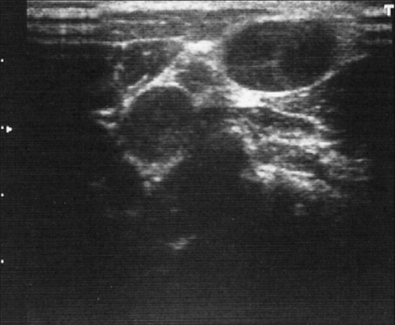
Well-defined, hypoechoic, oval lesions in cervical nodes. Echogenic thin layer is absent and margins are regular, suggestive of either metastasis or malignant lymphoma

Well-defined, hypoechoic, oval lesions in cervical nodes. Echogenic thin layer is absent and margins are regular, suggestive of either metastasis or malignant lymphoma