Figure 2.
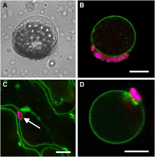
Tonoplast localization of the AtERDL6-GFP fusion protein in Arabidopsis and tobacco. Arabidopsis protoplasts (A and B) were transformed with an AtERDL6-GFP fusion construct. After 20 h of incubation, protoplasts were lysed by mild osmotic shock and analyzed by confocal laser scanning microscopy. A, Bright-field image of a protoplast after lysis, leaving the intact vacuole with attached chloroplasts. B, Fluorescence image of a lysed protoplast showing AtERDL6-GFP localization to the tonoplast (green) and chlorophyll autofluorescence (magenta) of attached chloroplasts. Tobacco leaves (C and D) were infiltrated with an Agrobacterium culture harboring the AtERDL6-GFP fusion construct pGP-67-1. After 2 d, infiltrated leaves were analyzed by confocal laser scanning microscopy. C, Epidermal leaf area depicting GFP fluorescence in tonoplasts of neighboring cells. D, Fluorescence image of a tobacco leaf protoplast after enzymatic degradation of the cell wall and lysis by mild osmotic shock, with GFP fluorescence restricted to the membrane of the remaining intact vacuole. Bars = 10 μm.

