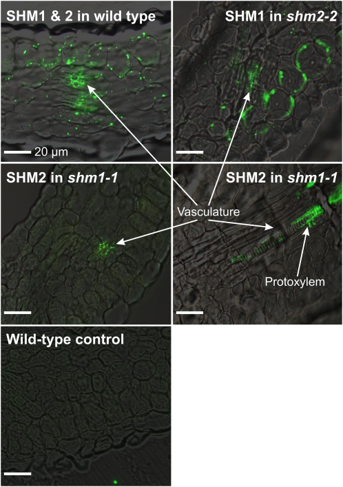
Figure 4.
Overlays of immunofluorescence signals of SHM1 and SHM2 and bright-field images of Arabidopsis leaf cross sections. Top left, SHM1 and SHM2 signals in mitochondria of the mesophyll and the vasculature of wild-type leaves. Top right, SHM1 signals in mitochondria of the mesophyll and the vasculature of shm2-2 leaves. Middle, SHM2 signals in mitochondria of the vasculature of shm1-1 leaves showing a likely location of SHM2 in the protoxylem. Bottom, Example of a control section subsequently treated with 5% bovine serum albumin instead of anti-SHM antibodies and the labeled secondary antibody. The primary antibody is specific for mitochondrial SHM and was affinity-purified against SHM2. The secondary antibody is conjugated with Alexa-Fluor 488. Bars = 20 μm.

