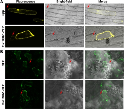
Figure 3.
Subcellular localization of OsTRXh1. A, Onion epidermal cells were transformed by particle bombardment. YFP (top panels) and OsTRXh1-YFP (bottom panels) fluorescence was observed with a confocal microscope. The onion epidermal cells were treated with 0.9 m mannitol for plasmolysis. B, The OsTRXh1-GFP fusion protein was secreted in the cell wall in N. benthamiana leaves. The top panels show the empty vector control, and the bottom panels show OsTRXh1-GFP fluorescence. Leaf epidermal cells were treated with 1 m sodium chloride for plasmolysis. Fluorescence, bright field, and merged images are shown for each transformation condition. Arrows indicate the cell wall.

