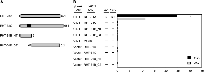
Figure 5.
Interaction between wild-type and mutant forms of RHT-B1 and TaGID1 in yeast two-hybrid assays. A, Schematic diagram showing full-length and mutant forms of RHT-B1, which were expressed as LexA activation domain (AD) fusions in yeast. Numbers indicate the amino acid positions within RHT-B1. The black box in the RHT-B1C schematic diagram indicates the position of the 30-amino acid insertion. The full-length TaGID1 was expressed as a LexA DNA-binding domain (DB) fusion protein in yeast. B, Interactions between activation domain and DNA-binding domain protein fusions were determined in cotransformed L40 yeast cells by scoring growth on His− medium containing 3-aminotriazole (3-AT; 0, 2, 5, 10, 30, and 60 mm). Data presented are maximum concentrations of 3-AT at which growth was observed; dashes signify no growth on 2 mm 3-AT. Quantitative values for each interaction were determined by measuring β-Gal activity in the presence and absence of 100 μm GA3. Values are means of three biological replicates ± se.

