Figure 2.
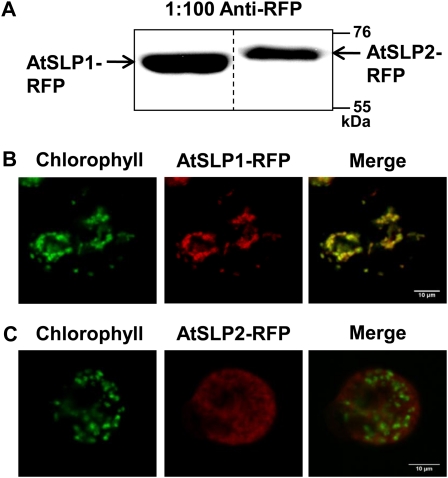
In vivo subcellular localization of AtSLP1 and AtSLP2 using stably transfected Arabidopsis cell culture. A, Western-blot verification of stably transfected cell culture constitutively expressing AtSLP1-RFP or AtSLP2-RFP. Each lane contains 30 μg of crude cell culture lysate probed with 1:100 anti-RFP IgG (Chromotek). B and C, Fluorescence images of protoplasts derived from stably transfected Arabidopsis cell culture expressing AtSLP1-RFP or AtSLP2-RFP (red), respectively. Chlorophyll autofluorescence is also shown (green), and images were merged to reveal localization. All images are single slices obtained using a confocal laser scanning microscope (Leica). Bars = 10 μm.

