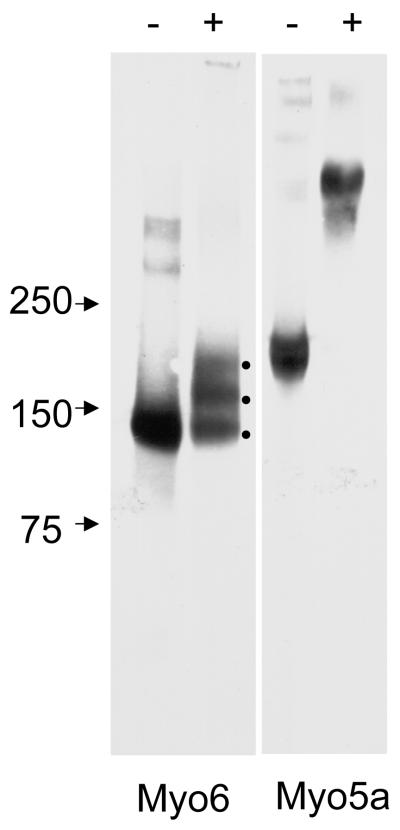
Fig. 1.
EDC crosslinking of human Myo6 and Myo5a. Immunoblot analysis of 25 μg/ml Myo6 and Myo5a without (-) and with (+) EDC crosslinking. Note the three bands in the Myo6 lane after EDC treatment presumably representing Myo6 heavy chain with 0-2 crosslinked CaM light chains. In contrast EDC treatment of Myo5a results in complete loss of monomeric heavy chain and a very high molecular weight smear is present, presumably representing Myo5a heavy chain dimer and variable numbers of light chains.

