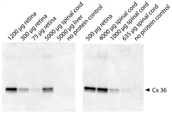
Figure 1.

Immunoprecipitation followed by western blot was used to quantify the amount of Cx-36 in spinal cord. Left panel shows the specificity of the antibodies. A band was detected at the correct size in retina and spinal cord lysates but not liver lysates. Right panel shows the sensitivity of the procedure. A band was detected using 1000 μg protein but was less clear with 635 μg protein. 800 μg was chosen for the study. Left and right panels show titration of retina and spinal cord protein to verify that the amount of antibody used was within the linear range for this procedure.
