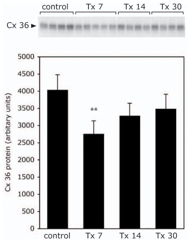
Figure 4.

Transient decrease below the level of the lesion in Cx-36 protein levels after Tx. Upper panel, Cx-36 western blot following immunoprecipitation from spinal cord from control rats or 7, 14 or 30 days after Tx (Tx 7, Tx 14, or Tx 30). Lower panel, quantification of the data shown in the upper panel. Note that Cx-36 protein level decreased at 7 days, ** denotes p>0.01.
