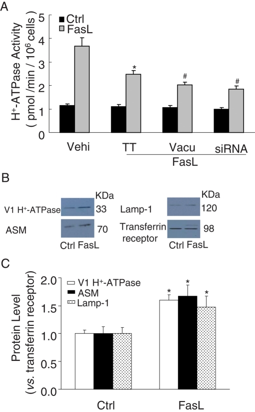
FIGURE 10:
H+-ATPase activity and protein expression on the cell membrane of bovine CAECs. (A) FasL (10 ng/ml) dramatically enhanced the activity of H+-ATPase on the cell membrane of CAECs by fluorometry, which was inhibited by pretreatment of these cells with TT (10 nM), vacuolin-1 (10 μM), or V1 H+-ATPase siRNA (n = 6, *p 0.05 vs. control; #p < 0.05 vs. only FasL-treated group). (B) Cell surface biotinylation assay for H+-ATPase subunit of V1 sector protein expression in bovine CAECs. Western blot gel document presents the relative levels of V1 H+-ATPase, ASM, lamp-1, or transferrin receptor on the cell membrane of CAECs. (C) Summary of results showing that the intensity ratio of V1 H+-ATPase to transferrin receptor increased by 59.5% on the cell membrane in response to FasL (10 ng/ml) treatment. The intensity ratio of ASM or Lamp-1 to transferrin receptor increased by 67.1 or 47.7%, respectively, on the cell member (n = 3, *p < 0.05 vs. control).

