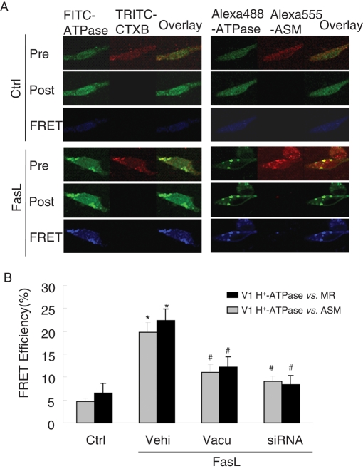
FIGURE 9:
FRET analysis of the MR marker ganglioside GM1, H+-ATPase, and ASM in bovine CAECs. FRET was detected using an acceptor-bleaching protocol. The blue images (representing FRET) on the bottom were obtained by subtracting a prebleaching image from a postbleaching image. (A) Representative images of FRET between V1 H+-ATPase and GM1 (CtxB labeling) or ASM. (B) Summarized results of detected FRET efficiency show that FasL significantly increased the FRET efficiency between V1 H+-ATPase and GM1 or ASM, which was effectively inhibited by vacuolin-1(10 μM) or V1 H+-ATPase siRNA (n = 6, *p < 0.05 vs. control; #p < 0.05 vs. only FasL-treated group).

