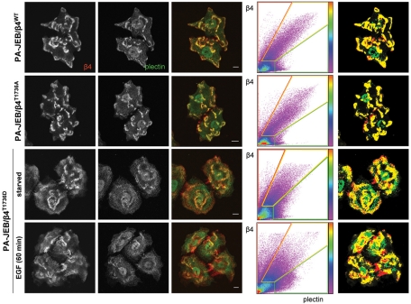
FIGURE 7:
The assembly of HDs in PA-JEB/β4 keratinocytes is prevented by mimicking the phosphorylation of β4 at T1736, which contributes to the EGF-induced HD disassembly. PA-JEB keratinocytes expressing wild-type or mutant β4 in which T1736 is substituted by alanine or aspartic acid were starved overnight, stimulated with or without 50 ng/ml EGF for 60 min, and fixed for immunolabeling of β4 (red) and plectin (green). The degree of colocalization of β4 and plectin is visualized in the overlay image (yellow) and using a scatter plot in which the intensity of β4 (y-axis) and plectin (x-axis) for each pixel is plotted. The color code is a measure for the number of pixels with similar β4/plectin intensity. Right, pixels in which β4 and plectin are strongly colocalized (yellow) and β4 (red) and plectin (green) are not colocalized.

