FIGURE 1:
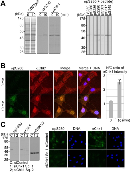
The specificity of anti–phospho-Ser-280 on Chk1 (αpS280) or anti-Chk1 (αChk1). (A) After 48 h of serum starvation, RPE1 cells were incubated with the fresh growing medium for 0 or 10 min as described in Materials and Methods. Specificity of each antibody was analyzed by immunoblotting. Amounts of loading cell lysates were determined by staining of SDS–PAGE gel with Coomassie brilliant blue (CBB; left). In the competition assay, αpS280 was preincubated with 50 ng/ml nonphosphopeptide S280 or each phosphopeptide (pS), and then the serum-stimulated cell lysate was immunoblotted (right). (B) Cells were stained with αpS280 (green), αChk1 (red), and 4′,6-diamidino-2-phenylindole (DAPI; blue; left). The N/C ratio of αChk1 intensity is shown. Data represent mean ± SEM for at least 20 cells in each cell group, **p < 0.01 using Student's t test (right). (C) RPE1 cells were transfected with the mixture of the indicated siRNA and Lipofectamine RNAiMAX reagent according to the reverse transfection procedures (Invitrogen). At 16 h after the transfection, the cells were cultured in the serum-free medium for 48 h and then incubated in the growing medium for 10 min. Similar diminishment of the antibody signals in the immunocytochemistry was observed in cells transfected with siChk1 Sq. 2 (unpublished data). Scale bar, 10 μm (B, C).

