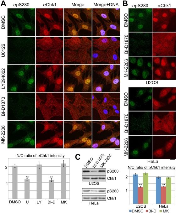
FIGURE 4:
Chk1 is translocated from cytoplasm to nucleus in a p90 RSK–dependent manner. RPE1 (A), U2OS, or HeLa (B, C) cells were treated with each chemical agent as described in Materials and Methods. At 5 (A) or 10 (B) min after serum addition, cells were stained with αpS280 (green), αChk1 (red), and DAPI (blue). In C, treated cells were also analyzed by immunoblotting. The N/C ratio of αChk1 intensity is also shown. Data represent mean ± SEM for at least 20 cells in each cell group, **p < 0.01 vs. cells treated with DMSO (A, B). Scale bar, 10 μm (A, B).

