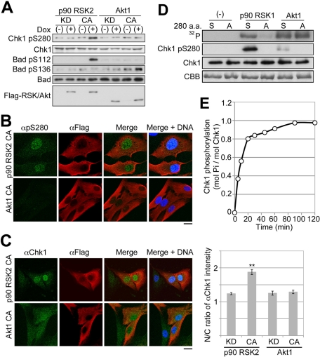
FIGURE 5:
P90 RSK phosphorylates Ser-280 on Chk1. (A–C) Tet-On RPE1 cell line was cultured in the serum-free medium for 48 h. After treatment, cells were cultured in serum-free medium with (+) or without (−) Dox for 6 h. Cells were analyzed by immunoblotting (A) or immunocytochemistry (B, C). CA or KD indicates a constitutively active or kinase-dead mutant, respectively. The N/C ratio of αChk1 intensity is shown. Data represent mean ± SEM for at least 20 cells in each cell group, **p < 0.01 vs. RSK2 KD-expressing cells (C; also see Supplemental Figure S1). Bar, 10 μm (B, C). (D) Purified Chk1 mutant KD (S) or KD/S280A (A) was incubated with or without p90 RSK1 or Akt1 for 20 min as described in Materials and Methods. The reaction mixture was analyzed by SDS–PAGE or immunoblotting with αpS280 (Chk1–pSer-280) or αChk1 (Chk1). After SDS–PAGE, bands of Chk1 or radioactive Chk1 were visualized by staining with CBB or autoradiography (32P), respectively. (E) Time course of Chk1 KD phosphorylation by p90 RSK1.

