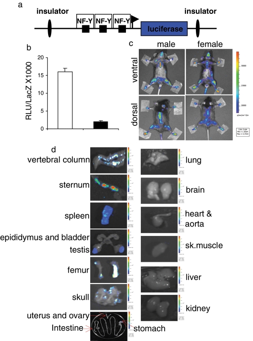
FIGURE 1:
NF-Y–dependent luciferase transcription in MITO-Luc mice. (a) Scheme of the transgene used. (b) C2C12 cells transiently transfected with the wild-type (white bar) or mutated (black bar) CCAAT (CCAAT versus TTACT) boxes. Transfection efficiency was normalized with cotransfected β-galactosidase expression vector (pCMV-lacZ). The error bars indicate the deviation of the mean of two experiments performed in triplicate. (c) BLI of representative MITO-Luc mice. The images were collected on 31 animals of each gender; one representative animal is shown. (d) Ex vivo BLI of positive and negative luciferase organs. After the animals were killed, the indicated organs were collected. Images of five animals for each gender were collected and one representative animal is shown. (c and d) Light emitted from the animal appears in pseudocolor scaling.

