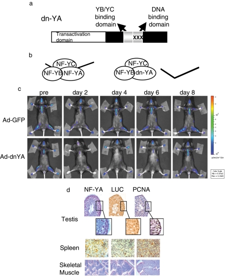
FIGURE 2:
NF-Y–dependent luciferase transcription in MITO-Luc mice. (a) Scheme of the dominant-negative NF-YA protein (dn-YA). (b) The NF-Y subunits, A, B, and C, form a complex that binds DNA. dn-YA is still able to interact with the NF-YB/-YC dimer, but the resulting trimer is inactive in terms of DNA binding. (c) BLI of representative MITO-Luc mice before (pre) and after (days 2, 4, 6, 8) injection of an adenovirus vector expressing GFP (Ad-GFP) or Ad-dnYA. The experiments were conducted on nine animals per group. Light emitted from the animals appears in pseudocolor scaling. (d) Immunohistochemical analysis of NF-YA, luciferase (LUC), and PCNA in testis (40×; insets: 100×), spleen (100×), and skeletal muscle (60×) tissues from a representative MITO-Luc mouse.

