FIGURE 1:
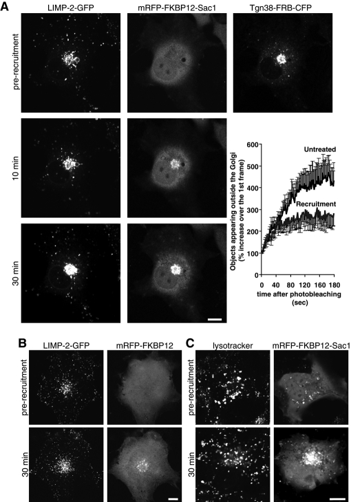
The effect of PtdIns4P elimination at the Golgi on LIMP-2 distribution in COS-7 cells. Cells coexpressing either mRFP-FKBP12-Sac1 (A) or mRFP-FKBP12 (B) together with Tgn38-FRB-CFP and LIMP-2-GFP were mounted on the microscope's heated stage (35°C) and treated with 100 nM rapamycin to induce recruitment of the cytosolic-FKBP12 constructs to the Golgi. Time-lapse images of individual cells were recorded for 30 min, and representative images are shown at 0 min (prior to recruitment) and at 10 and 30 min after recruitment. Graph shows morphometric analysis of the recordings in (A) using ImageJ software. The curves represent the number of individual fluorescent vesicles appearing outside the Golgi 15 min after the addition of rapamycin and photobleaching the whole cell outside of the Golgi area. The graph illustrates the first 200 s of the recordings taken at 0.5 frames/s. Each value represents a mean ± SEM of five recordings for each sample (control and rapamycin-treated). (C) Cells expressing mRFP-FKBP12-Sac1 and Tgn38-FRB-CFP were preincubated for 10 min with 100 nM LysoTracker Green at 35°C prior to addition of rapamycin. Scale bars: 10 μm.

