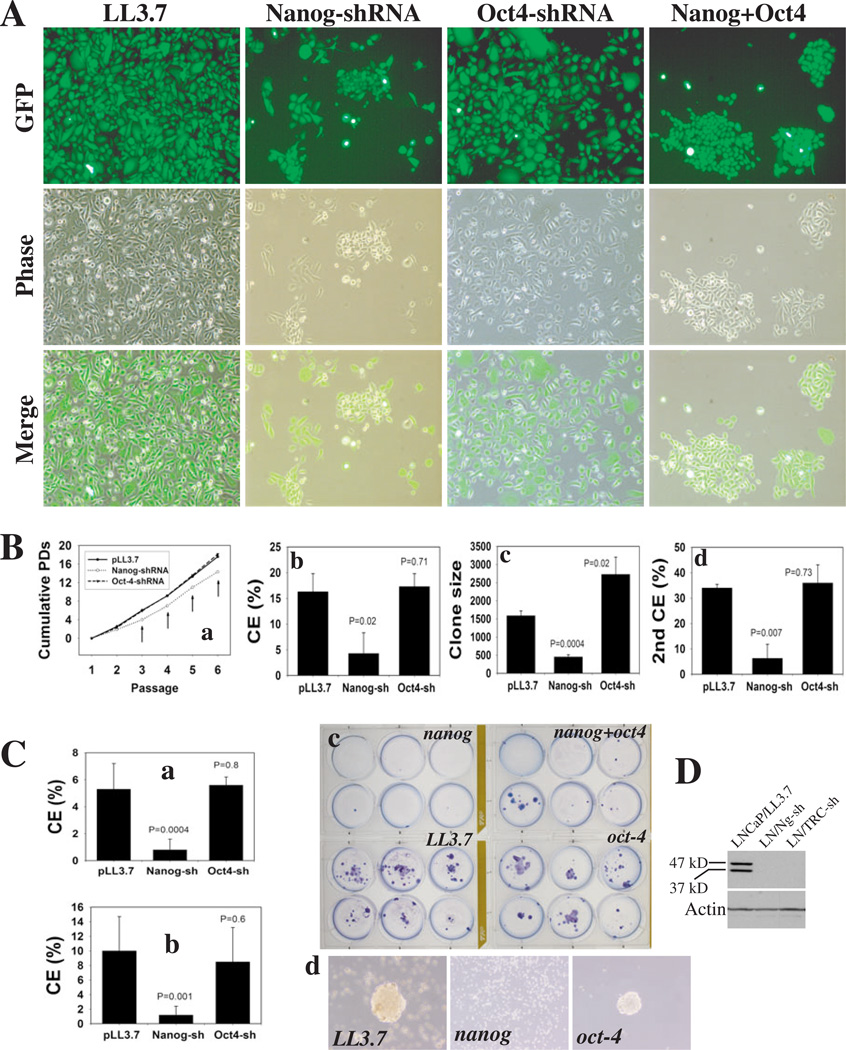
Figure 4. Nanog downregulation restricts clonal and clonogenic growth of PCa cells.
(A) PC3 cells infected with the indicated lentiviral vectors (MOI 25) were dissociated and replated, 72 h later, at clonal density in a 6-well plate (100 cells/well). Images 12 d post-plating (×200).
(B) PC3 cells infected with the indicated vectors (above) were serially passaged and cumulative PDs determined (a). Arrows, P<0.05. (b) 1o clonal efficiency (CE): 100 infected PC3 cells were plated per well in a 6-well plate; clones were counted 10 d or (c) 17 d after plating. (d) 2o clonal analysis: 1o clones were cloned out using a cloning ring and replated at 100 cell/well; 2o clones were counted at 10 d. For b-d, data for each condition were derived from at least 6 samples (mean ± S.D).
(C) 100 infected LNCaP cells (transduced as described above) were plated for CE and clones scored at (a) 14 d or (b) 37 d post plating. (c) Giemsa staining of clones at 37 d. (d) LNCaP 2o clonogenicity: 1,000 infected LNCaP cells were plated on 1% agarose, and, 15 d later, 1o spheres were harvested, dissociated with trypsin and replated.
(D) Western blotting (60 µg wcl/lane; eBio mAb) of LNCaP cells infected with LL3.7, Nanog- or TRC-shRNAs (72 h post infection).

