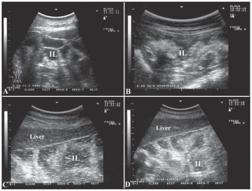
Figure 1.
Ultrasonographic appearance of the intestines in a healthy camel (A) and in 3 diseased camels (B, C, D) with Johne’s disease. Excessive accumulation of anechoic fluid, moderate to severe thickening and corrugation of the intestinal mucosa are apparent in sick animals. Images were taken at the right ventral abdomen using a 3.5 MH sector transducer. IL — intestinal loops.

