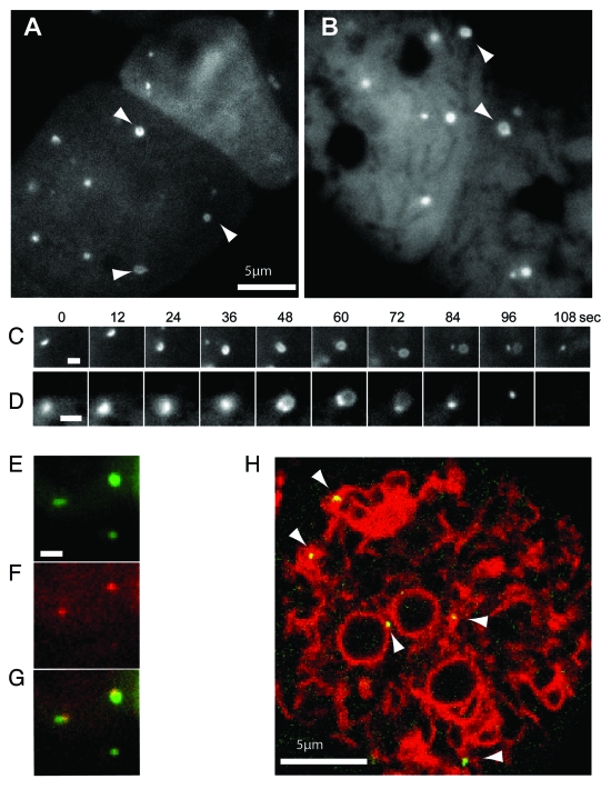
Figure 3.
High magnification imaging of the expansion of mechanically-induced autophagosomes. Ax3 cells expressing either (A) GFP-Atg8 or (B) GFP-Atg18 were imaged 20 min after capillary action-compression. Arrows indicate cup or circle structures, the full time-lapse series are shown in Video S2 and S3. Sequential images of individual forming autophagosomes visualized with GFP-Atg8 or GFP-Atg18 are shown in (C and D) respectively. Bars in (A and B) represent 10 μm and 2 μm respectively. Both markers localize to the same structure in compressed cells co-expressing (E) GFP-Atg8 and (F) tagRFP-Atg18; (G) shows the merged image and the complete time-lapse is shown in Video S1. (H) Image from a time-lapse series of compressed cells co-expressing the ER marker vmp1-mRFPmars and GFP-Atg18 observed by confocal microscopy. Arrows indicate forming autophagosomes. The full sequence can be seen in Video S4.

