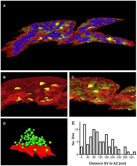
Figure 5. 3D reconstruction of a calyx of Held segment based on SEM images obtained from 100 consecutive sections.
A: Full reconstruction of a 31.7 µm3 calyx segment. The innervation side is orientated to the front. Red: plasma membrane; yellow: postsynaptic densities; blue: mitochondria; green: synaptic vesicles. B: Close up showing only plasma membrane and postsynaptic densities (colours as in A). C: Close up showing plasma membrane, postsynaptic densities and synaptic vesicles (colours as in A). D: 3D reconstruction of a single release site (colours as in A). E: Histogram of the distribution of distances between the presynaptic membrane and the synaptic vesicle membranes based on the release site shown in D.

