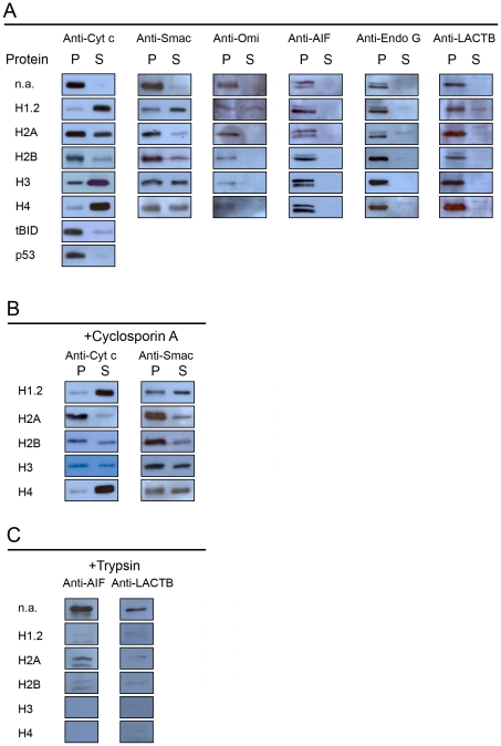
Figure 3. Release of mitochondrial intermembrane space proteins.
Mitochondria were incubated as described in the legend of fig. 1, whereupon they were sedimented by centrifugation and proteins of the resulting pellets (P) and supernatants (S) were analyzed by immunoblotting. Panel A: release of cytochrome c, Smac/DIABLO, Omi/HtrA2, AIF, endonuclease G, and LACTB by histones, tBID and p53. The concentrations used were: 5 µM histone H1.2; 10 µM histone H2A, 10 µM histone H2B, 5 µM histone H3, 5 µM histone H4, 1 µM tBID, and 1 µM p53. Panel B: effect of cyclosporin A on the histone-induced release of cytochrome c and Smac/DIABLO. Panel C: accessibility of AIF and LACTB to trypsin hydrolysis in the presence of histones.

