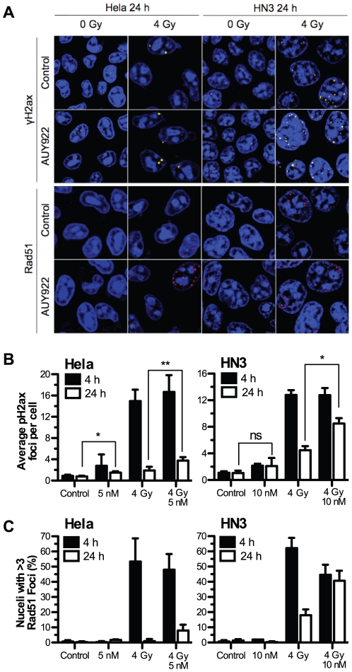
Figure 4. NVP-AUY922 delays radiation induced Rad51 foci formation and DNA damage repair.
HeLa and HN3 cells were plated in glass bottom dishes and after attachment exposed to NVP-AUY922 or DMSO control. 24 h post drug-treatment cells were mock irradiated or irradiated with 4 Gy, 4 h and 24 h post-irradiation cells were fixed and stained for dsDNA breaks using anti-phospho-H2ax and Rad51 focal formation with TO-PRO-3 as nuclear counter stain. (A) Representative images of remaining foci at 24 h are shown for HeLa and HN3 cells, area shown represents 60 µm by 60 µm. (B) Mean phospho-H2ax foci per-cell for both HeLa and HN3 at 4 h and 24 h post irradiation was quantified in three independent experiments consisting of phospho-H2ax counts for 150 cells per experiment. (C) Rad51 foci were quantified in two independent experiments with nuclei containing greater than 3 Rad51 foci scored as positive. Values stated are ± SEM for phospho-H2ax, ± SD for Rad51. Statistical analysis by student t-test between groups indicated, *p<0.05, **p<0.01.

