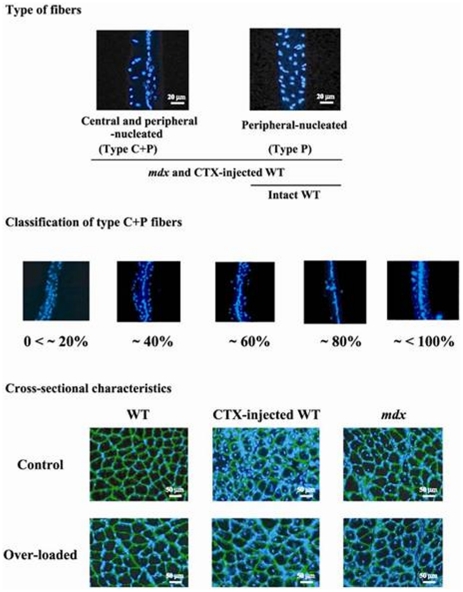
Figure 1. Representative cross-sectional and longitudinal characteristics of soleus muscle fibers in mdx and WT mice.
Immunohistochemical procedures were performed to stain myonuclei (blue) using Hoechst 33342 and laminin (green) using rabbit anti-laminin antibodies (Sigma, USA). mdx: dystrophin-deficient mice, WT: wild type mice, CTX: cardiotoxin, type P and C+P fibers: muscle fibers with myonuclear distribution at only peripheral and both central and peripheral regions, respectively. The characteristics of type C+P fibers were classified into 5 groups according to the percent distribution of central nuclei per single fiber; 0<∼20%, ∼40%, ∼60%, ∼80%, and ∼<100%. Greater differences were also noted in the fiber sizes of mdx mouse muscle.

