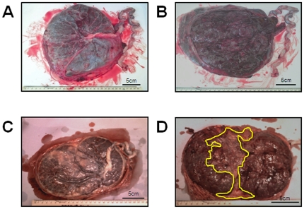
Figure 1. Examples of placental photographs from uncomplicated (A–B) and RFM (C–D) pregnancies.
A) Fetal side of a control placenta with a central cord insertion and normal distribution of chorionic plate vessels. B) Shows the maternal side of a control placenta. C) Shows the fetal side of a RFM placenta which is less round than the control example, and the cord is inserting laterally. D) Shows the maternal side of a RFM placenta. The area highlighted by the yellow line shows an abnormal white area which was confirmed on the cut surface of the placenta.

