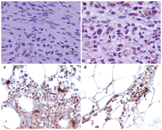
Fig. 3.
Immunohistochemical analysis of bone marrow of control and tumor-bearing hosts. a Cytoplasmic staining of NMT (arrow) in BMC from control rats. b Nuclear localization of NMT in BMC (short arrow) from a tumor-bearing rat (long arrows little cytoplasmic staining). c Mostly cytoplasmic NMT staining in bone marrow (BM) of control (arrows). d Intense nuclear (and some cytoplasmic) staining for NMT observed in the BM of a colon cancer patient (arrows) [adapted from Shrivastav et al. 2007]. Magnification: 20× dry (a, b), 10× dry (c, d)

