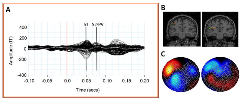
Figure 2.

Summary of MEG sensor data during tactile stimulation of the second digit (RD2). Averaged MEG sensor data (A) produces two peaks in primary somatosensory (S1) and secondary somatosensory/parietal ventral cortex (S2/PV). Equivalent current dipole fits (B) and sensor topography maps (C) of these sensor peaks place these sources in the contralateral (left) hemisphere.
