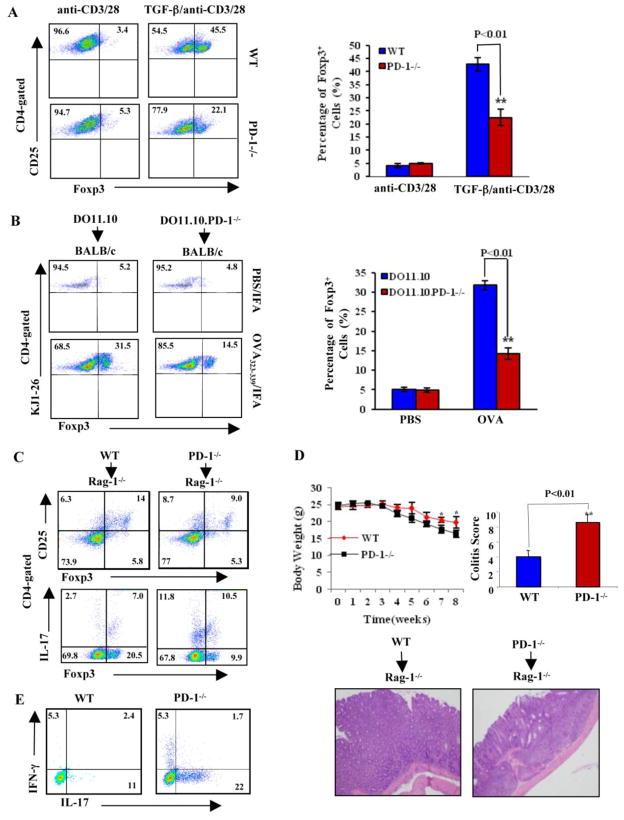
Figure 2. PD-1 potentiates iTreg generation in vitro and in vivo.
A. Naïve CD4+CD25− T cells from WT and PD-1−/− mice were cultured under iTreg differentiation protocol. The expression of Foxp3 was determined on CD4+CD25+ population (n=3). The figures are representative of three experiments repeated with similar results. B. CD4+KJ1-26+CD25− T cells from DO11.10 and DO11.10.PD-1−/− mice were adoptively transferred into BALB/c recipients, immunized with OVA323–339 in IFA by i.p, and Foxp3 expression in CD4+KJ1-26+ population was determined. The dot plot figures are representative of three experiments repeated with similar results. C. Rag-1−/− mice (n=5) were adoptively transferred i.v. with naïve WT or PD-1−/− CD4+CD25− T cells (5 × 10⁶/mouse). The expression of Foxp3, IFN-γ, and IL-17 within CD4+ T cells in draining lymph nodes collected from the recipients 8 weeks after transfer was determined by intracellular staining. D. The severity of colitis of the recipients was determined by body weight loss (upper panel) and H&E-stained paraffin sections of colons obtained 8 weeks after transfer of naïve CD4+CD25− T cells (lower panel). E. Naïve CD4+ T cells isolated from WT and PD-1−/− mice were stimulated under Th17 condition for 4 days. The cells were then collected and restimulated with PMA plus Ionomycin. IL-17-producing cells were determined by intracellular staining. These data are representative of at least four mice for each group, from three independent experiments with similar results.

