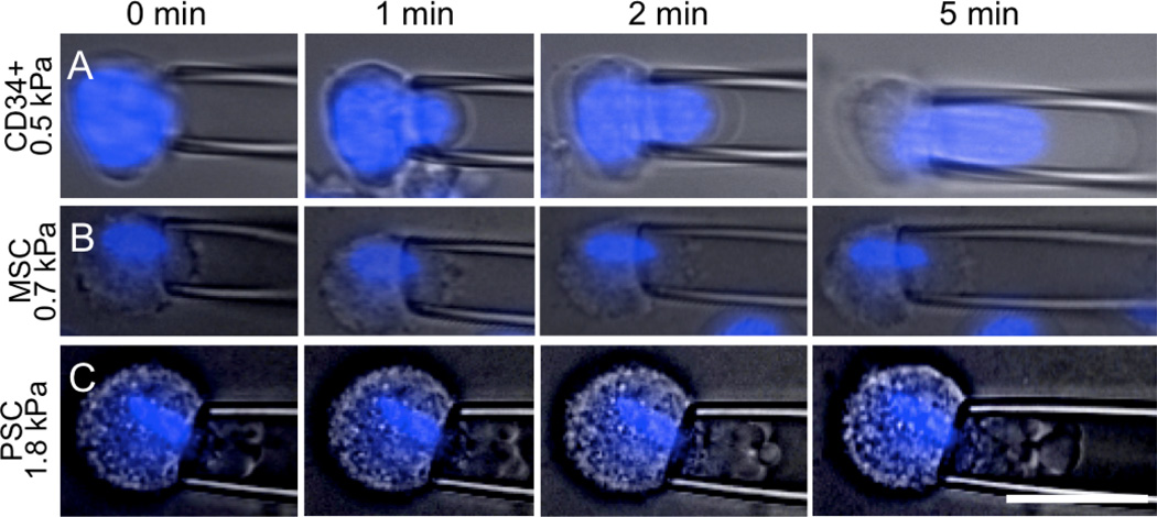
Figure 1. Micropipette aspiration of CD34+ cells, MSCs and PSCs.
Aspiration of the different cell types shows dramatically different response to force in magnitude of stiffness, viscoelastic deformation and contributions of cell components. Plasma membrane and nucleus (blue) deformations were followed over time after applying a constant aspiration pressure (in kPa, to the left of each set of images). (A) CD34+ cells showed significant viscous deformation with a strong contribution from the nucleus. (B) MSCs were less fluid, and showed slight contributions from a more condensed nucleus. (C) PSCs were stiff and showed minimal contributions from the nucleus. Scale is 25 µm.

