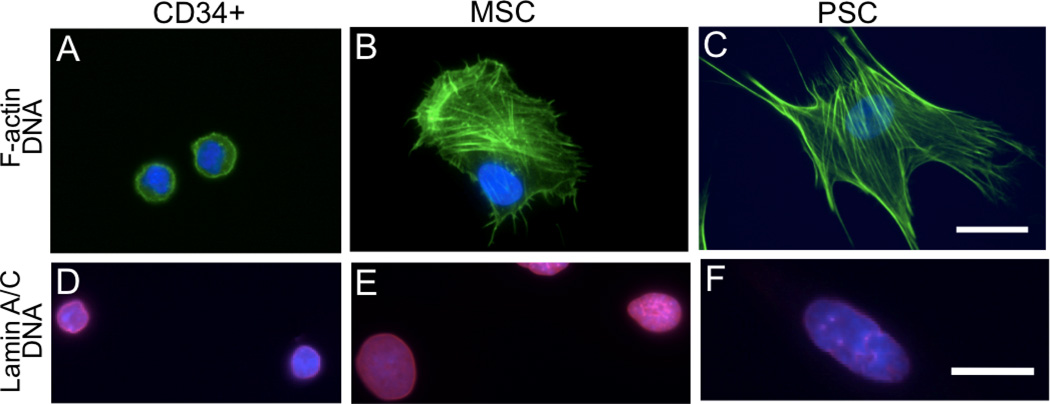
Figure 4. Actin, lamins and actin-nucleus connections are different among the studied cell types in adherent cultures.
(A) CD34+ cells show poorly developed F-actin (green) structures located around a central nucleus (blue), which occupies most of the cell volume. (B) MSCs show regions of higher development of F-actin stress fibers not coincident with nucleus. (C) PSCs show the most developed actin stress fibers colocalized with the nucleus. (D) A-type lamins (red) are homogeneously distributed in CD34+ cells, but are heterogeneous in MSCs (E) and PSCs (F). Scale is 25 µm; A–C and E–F scaled collectively.

