
Figure 7.
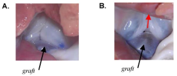
(A) Photograph looking up the left ventricular outflow tract at the isolated porcine aortic valve in which the noncoronary leaflet was replaced with a pericardium graft equal in size to the excised native leaflet (case W0H0). Photo was taken during application of 80 mmHg of air pressure. The valve was incompetent with a small central gap and regurgitant air flow. (B) Photograph looking up the left ventricular outflow tract at the incompetent isolated porcine aortic valve in which the noncoronary leaflet was replaced with a pericardium graft 25% wider than the excised native leaflet (case W25H0). Photo was taken during application of 80 mmHg of air pressure. Valve was incompetent, with a large (visible) central gap and substantial regurgitant air flow (direction indicated by red arrow).