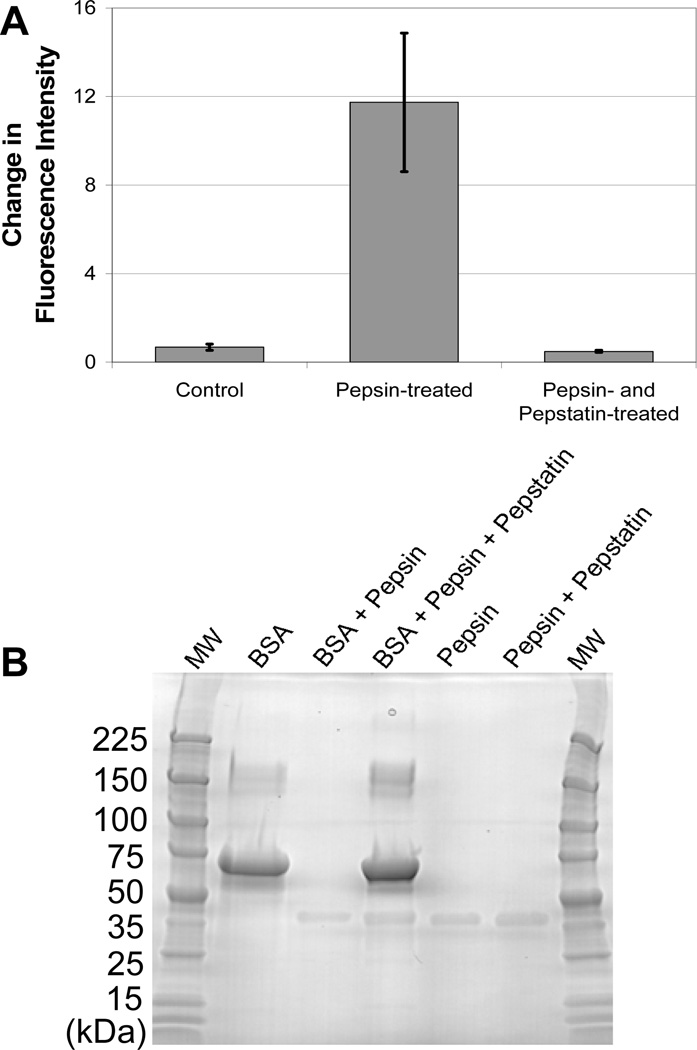
Fig. 2.
Dequenching of BSA is specific to protease activity. (A) Dequenching was not observed when BSA-AF647 was incubated with a combination of pepsin and pepstatin, a pepsin inhibitor. (B) Gel electrophoresis (4–20% gradient, polyarcrylamide, SimplyBlue SafeStain) shows that the degradation of BSA (67 kDa) by pepsin is inhibited by pepstatin. Pepsin (35 kDa) is visible on the gel while pepstatin (0.7 kDa) and the degraded BSA fragments are too small to observe.

