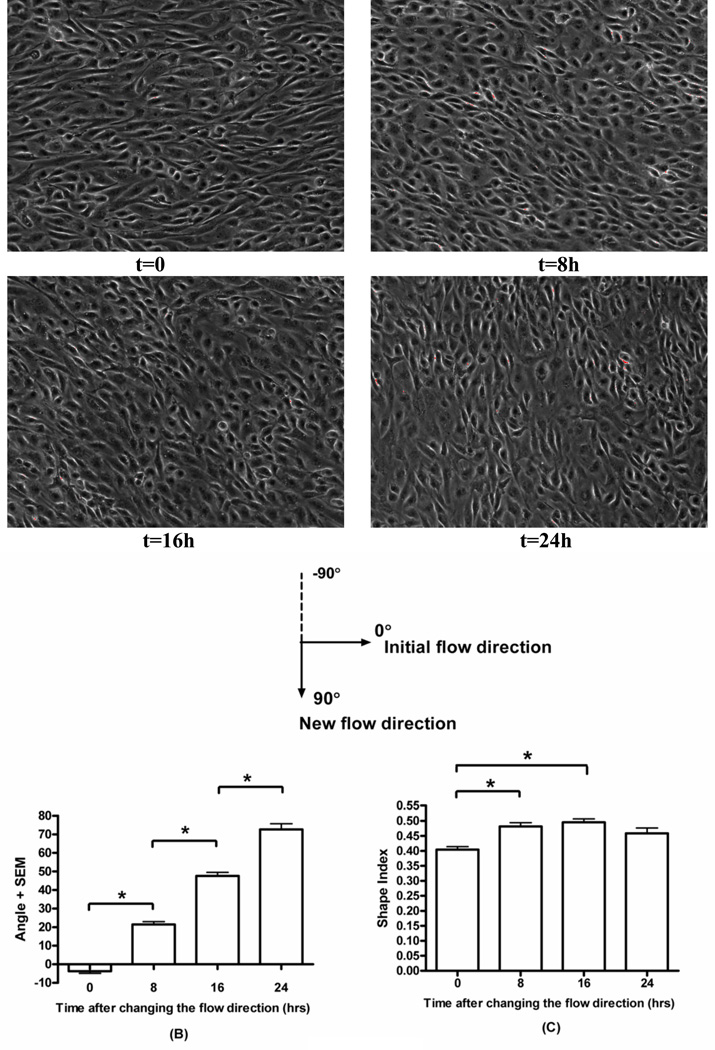
Figure 4.
Re-alignment of cell morphology. BAECs were pre-conditioned by shear (12 dynes/cm2, 24h). Initial flow direction was from left to right, then was changed to vertical (up to down). t indicates time after the direction change. A): Phase-contrast images of shear pre-conditioned endothelial cell monolayers exposed to the vertical flow for indicated times. B) Orientation angle as a function of time of exposure to vertical shear. Values are means ± SEM. (*P<0.001, n=3). C) Shape index as a function of time of exposure to the vertical flow. (*P<0.001, n=3).

