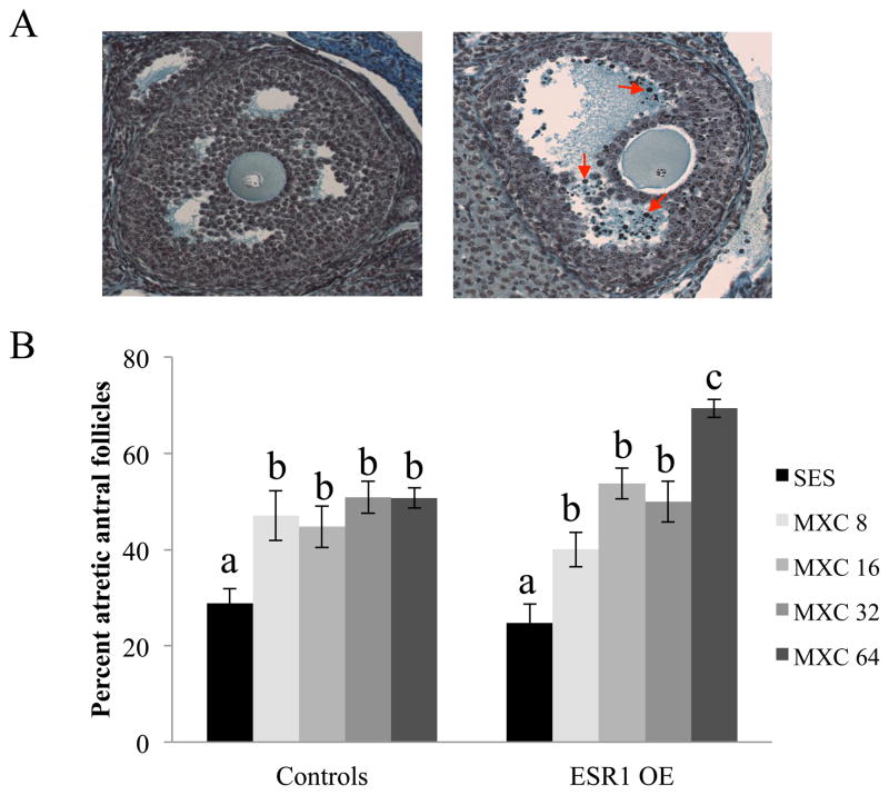
Figure 1.
Evaluation of atresia in control and ESR1 OE ovaries treated in vivo with sesame oil (SES) or MXC (8 – 64 mg/kg/day). Ovaries of mice were collected and fixed for histological evaluation of atresia. (A) Histological sections of ovaries were analyzed for atresia by comparing the presence of apoptotic bodies. Representative sections of a healthy antral follicle (left panel) and an atretic antral follicle (right panel) with red arrows pointing to a few apoptotic bodies are shown. (B) The percentages of atretic antral follicles were quantified and plotted on a graph. Each bar represents means ± SEM. Bars with different letters are significantly different from each other (n = 6; p≤0.05).

