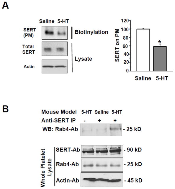
Fig. 3.
(A) Western blot analysis of SERT expression in platelets from saline- and 5-HT-infused mice. Representative images are from one of four independent experiments. Total SERT in platelet lysates was similar between platelets from the two groups of animals, but SERT was depleted in the platelet plasma membrane (PM) of 5-HT-infused mice. Actin was used as a loading control for each lysate. Average immunodensity values reveal that the density of SERT on the PM of platelets from 5-HT-infused mice was 42% less compared to control mice. * = statistical difference between saline- and 5-HT- infused mice. All assays were performed in triplicate (n = 15 group). (B) SERT-Rab4 association is enhanced in platelets from 5-HT-infused mice. Platelets were isolated from saline- and 5-HT-infused mice. Platelet lysates were incubated with an anti-SERT Ab (lanes 2 and 3) or pre-immune serum (lane 1). Proteins were eluted from PA beads and resolved on SDS-PAGE followed by WB using an anti-Rab4 Ab to determine Rab4-SERT association; or to determine the expression level of Rab4 in whole lysates. Expression of SERT in total platelet lysates was determined by WB. A distinct 25 kD band was only observed in platelets from 5-HT-infused mice. Rab-4 was not detected in anti-SERT immunoprecipitate using platelets from saline-infused mice. Actin levels were used as a sample loading controls.

