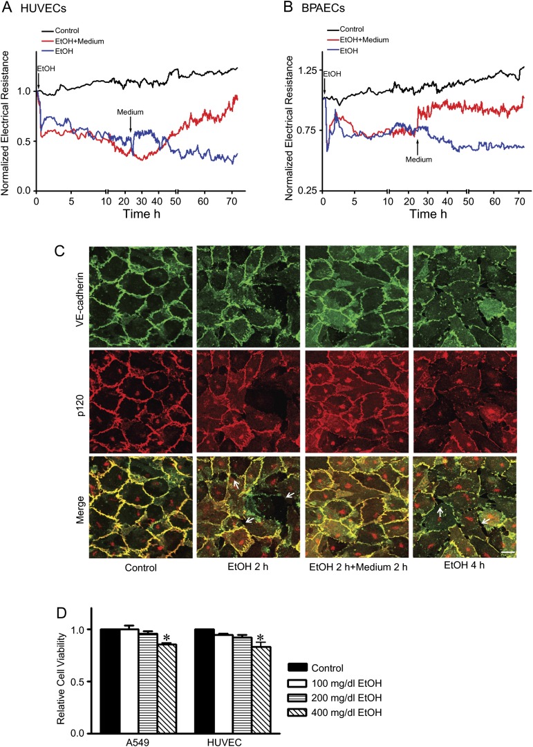
FIG. 2.
Reversibility of ethanol’s effect on endothelial cells. (A and B): HUVECs and BPAECs were exposed to ethanol (0 or 200 mg/dl) for 24 h. For some groups, the culture medium was removed at 24 h and replaced with fresh medium containing no ethanol, and cells were grown in this medium for an additional 48 h. Electrical resistances on HUVEC and BPAEC monolayer were continuously recorded. Arrows indicate the time of ethanol exposure and replacement of fresh medium. (C): HUVEC monolayer was exposed to ethanol for 2 h. For some groups, ethanol-containing medium was removed, and cells were washed and replaced with medium containing no ethanol for an additional 2 h. The expression of VE-cadherin and p120 was visualized as described in Figure 1. Arrows indicate the intercellular gaps. Scale bar = 20 μm. (D): A549 and HUVECs were exposed to ethanol (0, 100, 200, or 400 mg/dl) for 24 h, and cell viability was determined with MTT assay as described under the Materials and Methods section. The number of viable cells was presented relative to untreated controls. Each datum point was the mean ± SEM of three independent experiments. * denotes a statistically significant difference from untreated controls (p < 0.05).

