FIG. 7.
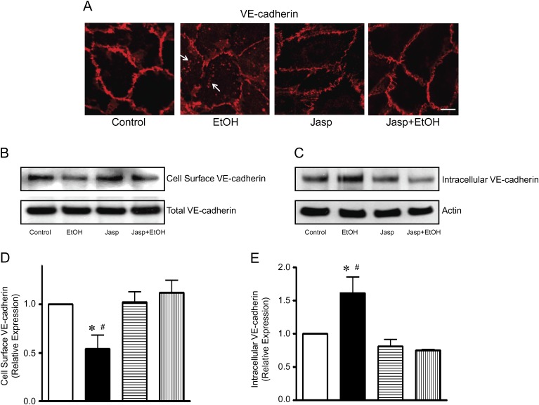
Effect of jasplakinolide on ethanol-induced endocytosis of VE-cadherin. (A) HUVEC monolayer was pretreated with jasplakinolide (Jasp, 50nM) for 4 h and then exposed to ethanol (0 or 200 mg/dl) for 4 h. VE-cadherin was visualized by immunofluorescence microscopy. Arrows indicate the cytoplasmic VE-cadherin. Scale bar = 10 μm. (B and C): HUVEC monolayer was pretreated with Jasp (50nM) for 4 h and then exposed to ethanol (0 or 200 mg/dl) for 6 h. The expression of cell surface VE-cadherin (B) and intracellular VE-cadherin (C) was determined as described in Figure. 5. (D and E): The relative expression of cell surface VE-cadherin (D) and intracellular VE-cadherin (E) was determined by densitometry and normalized to the total VE-cadherin (D) or actin (E), respectively. * denotes a statistically significant difference from untreated controls. # denotes a significant difference from jasplakinolide-treated groups (p < 0.05). These experiments were replicated three times.

