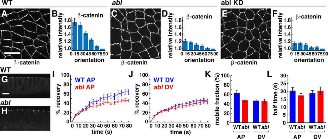
Figure 5. Abl is required for the planar polarized localization and dynamics of β-catenin.
(A–F) β-catenin localization in WT (A, B), abl mutant (C, D), and abl KD embryos (E,F). Values were normalized to the mean intensity of edges perpendicular (75°–90°) to the AP axis. β-catenin planar polarity was significantly reduced in abl mutant (p=0.0002) and abl KD embryos (p=0.000017) (n=7 WT, 12 abl mutant, 3 abl KD embryos, 76–169 edges/embryo). Anterior left, dorsal up. Cross sections shown in G,H. (I,J) The percentage of pre-bleach fluorescence observed after bleaching β-catenin:GFP at AP or DV junctions. (K) In WT, β-catenin displayed a higher mobile fraction at AP junctions (63+/−6%, n=19 edges) (mean+/−s.e.m.) than at DV junctions (47+/−4%, n=25) (p=0.023). This difference was abolished in abl mutants (48+/−3% at AP junctions, n=25, 46+/−4% at DV junctions, n=20) (p=0.317). (L) The time to recover half maximal fluorescence (t½) was similar for all edges. Bars, 10 µm. See also Figure S3.

